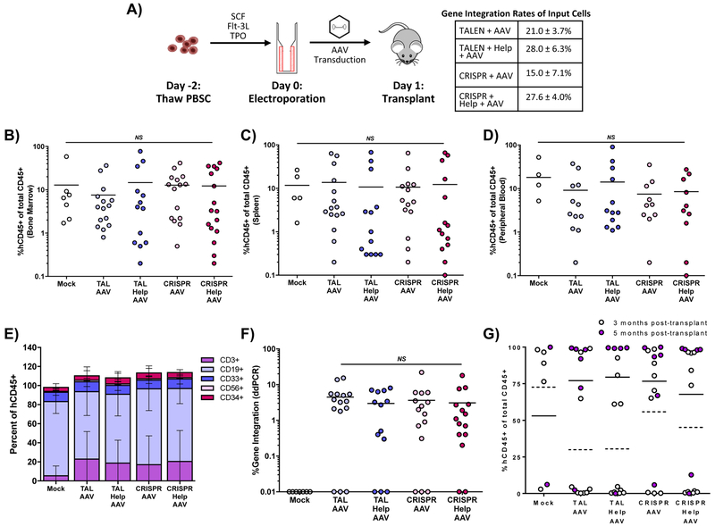
Figure 5. In vivo assay of CD34+ PBSC in NSG mice after treatment with TALEN or CRISPR, AAV6 CD40L cDNA donor and adenoviral helper proteins.
A) Schematic of NSG mouse transplants and gene integration of input cells as measured by ddPCR. Engraftment of gene modified human PBSC in B) bone marrow, C) spleen, and D) peripheral blood of transplanted mice, determined as the percentage of human CD45+ cells of all human and murine CD45+ cells. E) Lineage distribution of human cells engrafted in NSG mice in the bone marrow 12 weeks post-transplant using fluorescent-labeled antibodies to human T cells (CD3), human B cells (CD19), human myeloid cells (CD33), human NK cells (CD56), and human progenitors (CD34). F) Gene editing determined by ddPCR in bone marrow 12–20 weeks after transplant. G) Thymic engraftment analyzed at 3 and 5 months post-transplant. Data were analyzed by Wilcoxon Rank-Sum Test. **p≤0.01, NS = not significant. See also Figure S5.

