
Figure 1.
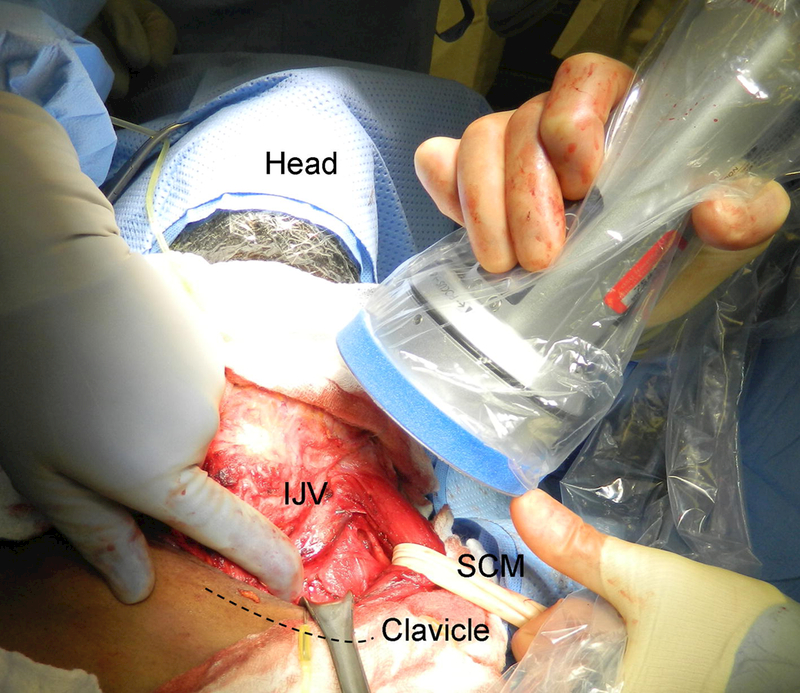
Position for imaging the thoracic duct. The probe is held at the patients head near the angle of the mandible and pointed caudally to image the space beneath the clavicle. The sternocleidomastoid muscle (SCM) soft tissue above the clavicle (dashed line), and internal jugular vein (IJV) are retracted to allow for better visualization.