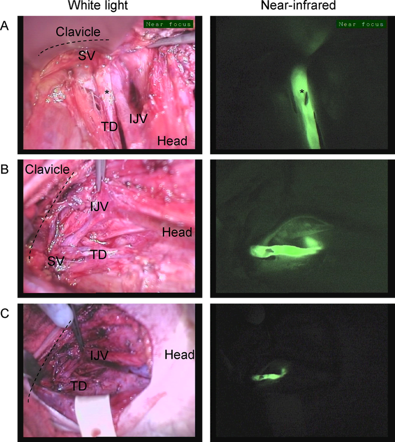
Figure 2.

Near-infrared imaging of the thoracic duct in Patient #4. The thoracic duct (TD) inserting on the junction between the Internal jugular vein (IJV) and Subclavian vein (SV) from 3 different points of view. The TD is marked with a clip (*) which is clearly visible on the near-infrared images A. Imaging from the angle of the mandible down to the space below the clavicle (dashed line), B. Imaging as the operator is standing on the left side of the patient C. Similar view as in B. but imaged from a farther distance from the patient.
