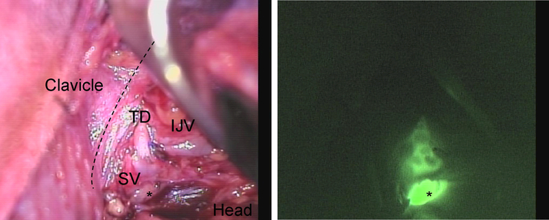
Figure 3.

Near-infrared imaging of the thoracic duct in Patient #5. The thoracic duct (TD) inserting on the junction between the Internal jugular vein (IJV) and Subclavian vein (SV) imaging from the head down to the space below the clavicle. A level IV lymph node (*) is also fluorescent.
