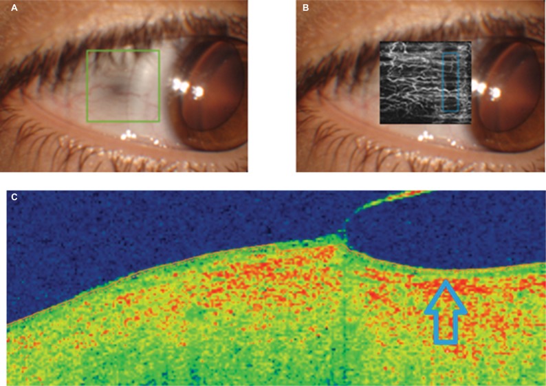
Figure 3.
OCT analysis of the peripheral bearing of the scleral contact lens with a sagittal height of 4800 µm (A, frontal image; C, OCT section showing the position of the peripheral area of the lens in a specific meridian), including the angiography OCT examination of the impact on the conjunctival vascular flow (B).
Note: Compression area of the peripheral edge of the scleral contact lens is indicated by the arrow.
Abbreviation: OCT, optical coherence tomography.

