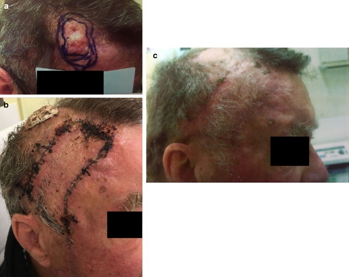
Fig. 1.
a Photograph of a biopsy-proven poorly differentiated squamous cell carcinoma with 5-mm excision margins marked. b 2-week postoperative photograph of the advancement flap with burrow triangle excision based on the frontal branch of the superficial temporal artery. c Photograph of flap and hair-bearing areas 6 weeks postoperatively

