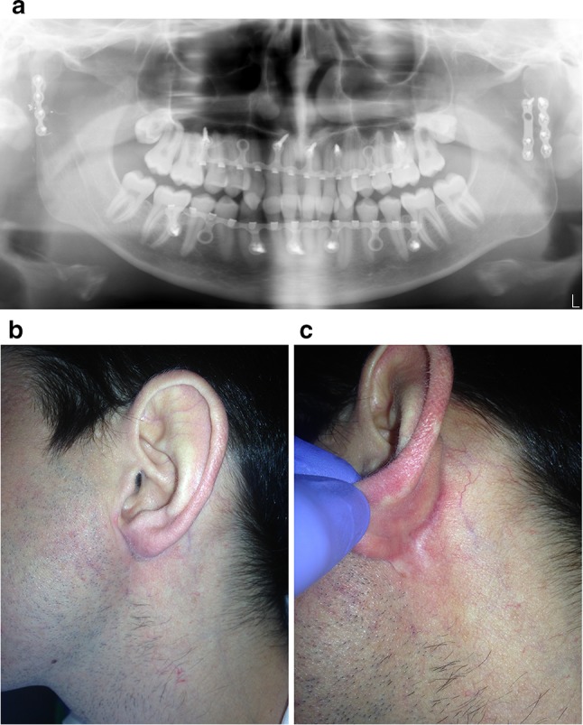
Fig. 3.

Case 1: a panoramic radiograph of the postoperative phase: the image shows the hybrid system used for intermaxillary fixation and to guide postoperative occlusion. Two 2.0-mm plates used for osteosynthesis in the left condyle and a 2.0-mm plate for osteosynthesis in the right condyle; b, c appearance of the scars 1 year postoperatively
