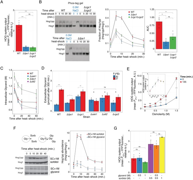
Figure 4.
Heat-shock activation of HOG involves efflux of glycerol. (A) HOG transcriptional reporter in WT, Δfps1 and Δrgc1Δrgc2 grown in SC + 1 M sorbitol measured 2 h after a shift from 30 °C to 37 °C. (B) Phos-tag gel showing HOG MAPK activation dynamics in WT, Δfps1 and Δrgc1Δrgc2 grown in SC + 1 M sorbitol following a shift from 30 °C to 37 °C, and a control of WT cells grown in SC stimulated with 0.5 M NaCl for 5 minutes. Left: representative blot. Middle. Fraction of phosphorylated Hog1. Right. Mean area under the curve (AUC) of phosphorylated Hog1 ± SEM. Uncropped blot in Fig. S11. (C) Intracellular glycerol measured in the indicated strains grown in 0.5 M NaCl after a shift from 26 °C to 37 °C. (D) Accumulated extracellular glycerol during 300 minutes of growth at 30 °C or after transfer to 37 °C of cells grown in SC + 1 M sorbitol. “a” p > 0.1 and “b” p < 0.05. (E) HOG transcriptional reporter in WT adapted to the indicated osmolarities at 30 °C and then transferred to 37 °C. Inset shows the increase in reporter due to the temperature shift relative to t0. (F) HOG MAPK activation dynamics in WT adapted to 1 M sorbitol or 1 M glycerol, grown at 30 °C and then transferred to 37 °C. Diagram. In medium with sorbitol, when Fps1 opens, glycerol will be lost from the cell. However, in medium with glycerol, even if Fps1 opens, the net glycerol flux should be zero. Left. Representative blot. Right. Phosphorylated Hog1 abundance. (G) HOG transcriptional reporter in WT adapted to the indicated mixtures of glycerol and sorbitol at 30 °C and then transferred to 37 °C. In all panels, data correspond to the mean of three independent replicates (shown as dots) ± SEM. Strains: LD3342, RB3396, RB3710, RB3717, RB3722, RB3376 and RB3382.

