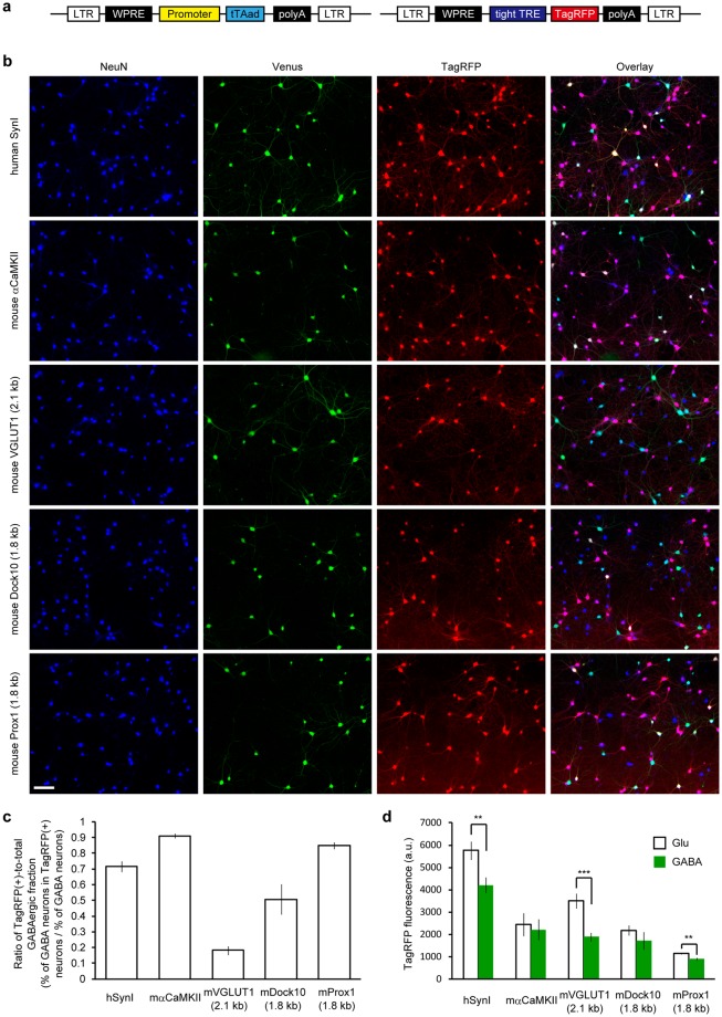
Figure 1.
Glutamatergic neuron-specificity of the five different lentiviral promoters assessed in cultured hippocampal neurons from VGAT-Venus Tg mice. (a) Schematic drawing of a pair of the lentiviral vectors that depend on the Tet-Off system to drive TagRFP expression under the promoters tested in this work. Transgene sequences flanked by long terminal repeat (LTR) sequences, which facilitate the integration into the host genome, are shown. A regulator vector (left) expresses an advanced tetracycline transactivator (tTAad) under the control of a given promoter and a response vector (right) expresses TagRFP in the presence of tTAad. See the Materials and Methods section for details. (b) Fluorescence images of cultured VGAT-Venus Tg neurons virally-expressing TagRFP using the five different promoters. Neuronal somata are indicated by anti-NeuN immunostaining (blue). Venus fluorescence, amplified by anti-EGFP immunostaining, indicates GABAergic neurons (green). TagRFP fluorescence indicates reporter expression (red). Note that TagRFP-positive GABAergic neurons, indicated by a white appearance in the merged images are rarely observed in the VGLUT1 promoter condition. Scale bar indicates 100 μm. (c) The ratio of TagRFP-positive populations within GABAergic neurons that was obtained by dividing the percentage of GABAergic neurons in the TagRFP-positive neurons by the percentage of total GABAergic neurons in the culture. A smaller value indicates a higher specificity towards glutamatergic neurons. The 2.1 kb of the mouse VGLUT1 promoter provided a significantly smaller ratio than all of the other promoters that were tested (p < 0.01, One-way ANOVA followed by Bonferroni’s test). The number of coverslips (n) analyzed in each group are represented in Table 1. (d) Quantification of TagRFP fluorescence intensity in neuronal soma. The synapsin I promoter, the VGLUT1 promoter and the Prox1 promoter provided a significantly higher expression in glutamatergic neurons than in GABAergic neurons (***p < 0.001, **p < 0.01, unpaired t-test).

