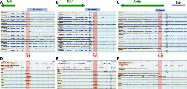
Figure 2.
Novel STEs demarcate chromatin domain boundaries and bind to insulator proteins. Top, screen crops of the repressive H3K9Me3 and H3K27Me3 ChIP-seq profiles surrounding STEs in lab (A), Dfd (B), and Antp (C) genomic regions from different embryonic stages. The EcoRI fragments captured by SF1 are indicated by the blue-shaded horizontal bars on top. The sub-fragments containing enhancer-blocking activities (En-B, also see Fig. 3) are indicated by the red shaded vertical bars with their names indicated below. Bottom, screen captures of ChIP-Chip profiles of known insulator proteins surrounding the STEs in 0–12 h embryos64. Yellow-shaded boxes represent called peaks for bound proteins. Map coordinate is based on ModEncode GBrowser dm3 (BDGP R5).

