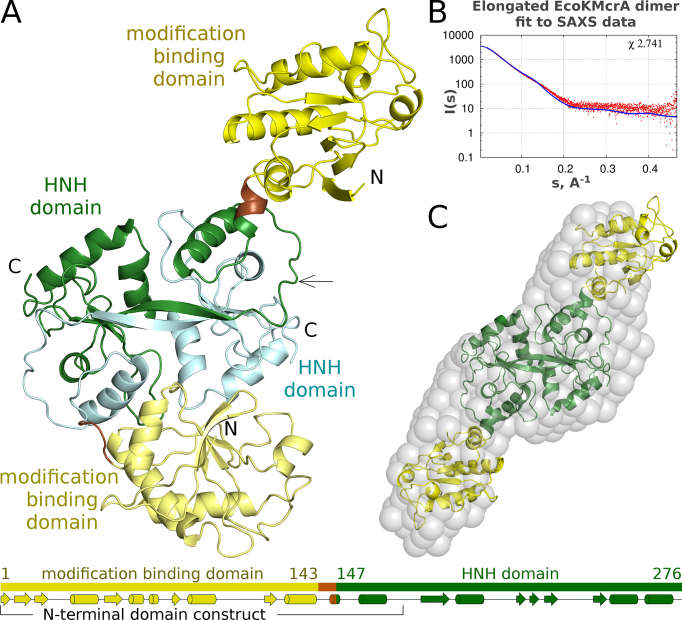
Figure 3.
Structure of EcoKMcrA. (A) Ribbon diagram of the EcoKMcrA dimer in the asymmetric unit of the crystals, colored according to domains. The domain organization of each protomer of is shown below. (B) Comparison of a symmetrized model of the EcoKMcrA dimer based on the more elongated protomer, with small-angle X-ray scattering (SAXS) data for the protein in the absence of DNA. (C) EcoKMcrA model ab initio calculated from the SAXS data (grey spheres) overlaid with the symmetrized dimer that was used for the calculation of the predicted small angle X-ray scattering data presented in panel B.

