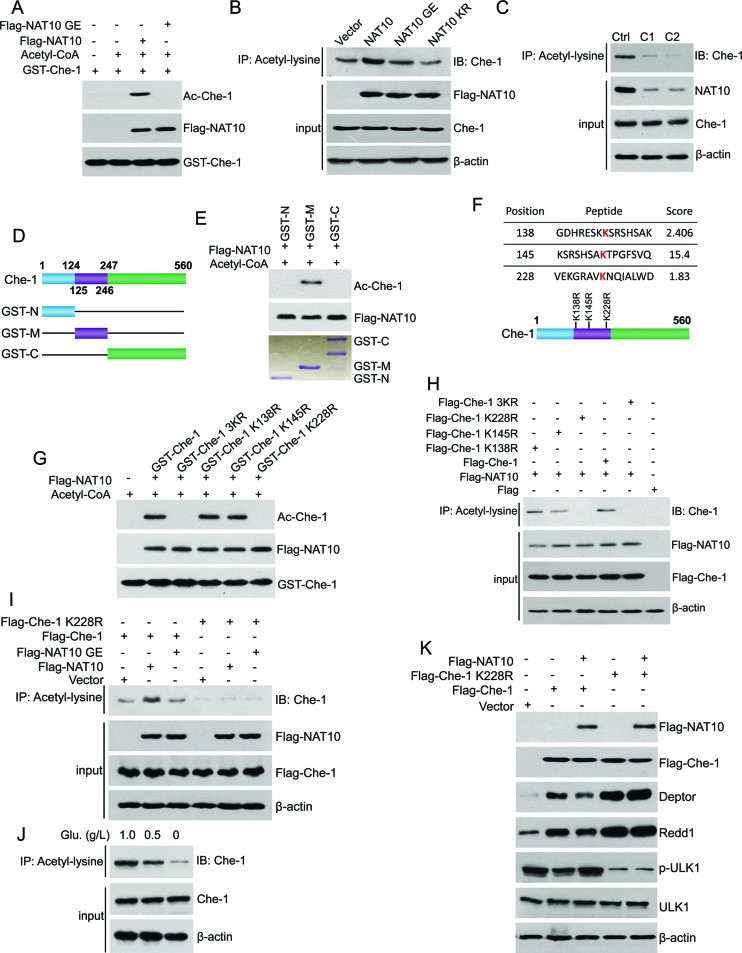
Figure 5.
NAT10 acetylates Che-1 at K228. (A) In vitro acetylation experiments were performed with GST-Che-1 and purified Flag-NAT10 or Flag-NAT10 GE as described in the Materials and Methods. Acetylation of Che-1 was evaluated by Western blot using an anti-acetyl-lysine antibody. (B) HCT116 cells were transfected with the indicated plasmids. Acetylation of Che-1 was evaluated by Western blot using an anti-Che-1 antibody on anti-acetyl-lysine antibody-specific immunoprecipitants. Inputs were evaluated by Western blot using the indicated antibodies. (C) Cell lysates were prepared from HCT116 Ctrl cells or HCT116 NAT10 KO cells (c1 or c2). Acetylation of Che-1 was evaluated by Western blot using an anti-Che-1 antibody on the anti-acetyl-lysine immunoprecipitants. The expression of NAT10 and Che-1 was evaluated by Western blot using the indicated antibodies. (D) The schematic diagram represents the Che-1 deletion mutant constructs. (E) In vitro acetylation experiments were performed with purified GST-Che-1-N, GST-Che-1-M or GST-Che-1-C fusion proteins and purified Flag-NAT10 as described in the Materials and Methods. Acetylation of Che-1 was evaluated by Western blot using an anti-acetyl-lysine antibody. (F) The potential acetylation sites on Che-1-M were predicted using the biocuckoo website. (G) In vitro acetylation experiments were performed with the purified GST-Che-1, GST-Che-1-3KR, GST-Che-1-K138R, GST-Che-1-K145R or GST-Che-1-K228R fusion protein and Flag-NAT10 as described in the Materials and Methods. Acetylation of Che-1 was evaluated by Western blot using an anti-acetyl-lysine antibody. (H) HCT116 cells were transfected with the indicated plasmids. Cells were harvested, and immunoprecipitation was performed to evaluate the level of Che-1 acetylation (upper). The expression levels of Flag-tagged proteins in the cell lysates were evaluated by Western blot using the indicated antibodies (lower). (I) U2OS cells were transfected with the indicated plasmids. Immunoprecipitation was performed using an anti-acetyl-lysine antibody to evaluate the level of Che-1 acetylation (upper). The expression levels of Flag-tagged proteins in the cell lysates were evaluated by Western blot using the indicated antibodies (lower). (J) HCT116 cells were cultured in medium containing different concentrations of glucose for 18 h. The acetylation levels of Che-1 in the anti-acetyl-lysine antibody-specific immunoprecipitants were evaluated using an anti-Che-1 antibody (upper). Total Che-1 levels were evaluated by western blot (lower). (K) HCT116 cells were transfected with the indicated plasmids. Cell lysates were prepared and subjected to Western blot for evaluation of the indicated proteins. See also Supplementary Figure S4.

