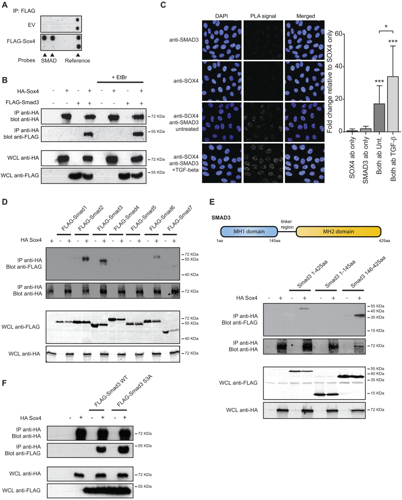
Figure 1.
SOX4 associates with the MH2-domain of SMAD3 independent of receptor-mediated phosphorylation. (A) MCF7 cells were transfected with FLAG-Sox4 or an empty vector control. Biotin-labeled transcription factor binding probes were added to the cell lysate after which Sox4 was immunoprecipitated using a FLAG- antibody. Subsequently, specifically bound probes were hybridized to the TF-TF array and visualized using streptavidin antibodies. (B) HEK293T cells were transfected with HA-Sox4 and FLAG-SMAD3. Lysates were treated with 25 μg/ml EtBr for 20 min after which Sox4 was immunoprecipitated and immunoblots were probed for HA and FLAG. Results are representative of at least three independent experiments. (C) The SOX4-SMAD3 interaction was analyzed by PLA. HMLE cells were treated with 2.5 ng/ml TGF-β overnight or left untreated. Left panel: Punctate staining indicates the specific interaction between the two proteins and DAPI was used to co-stain the nucleus. Right panel: Quantification of punctate staining relative to anti-SOX4 condition (negative control). Results are representative of three independent experiments. (D) HEK293T cells were transfected with HA-Sox4 and FLAG-SMAD1–7. Sox4 was immunoprecipitated from cell lysates and co-immunoprecipitation of SMAD proteins was assessed by immunoblot for HA and FLAG-epitope antibodies. Results are representative of at least three independent experiments. (E) Top panel: Schematic representation of SMAD3 protein. Bottom panel: HA-Sox4 was immunoprecipitated from HEK293T cells co-transfected with full-length SMAD3 (1–425aa), the N-terminal (1–145aa) or C-terminal SMAD3 regions (146-425aa). Results are representative of at least three independent experiments. (F) HEK293 cells were transfected with HA-Sox4 and Flag-SMAD3 wild-type (WT) or phosphorylation-defective SMAD3 S3A. Sox4 was immunoprecipitated from cell lysates and protein expression was assessed by immunoblot using anti-HA and Flag antibodies. Images are representative of three independent experiments.

