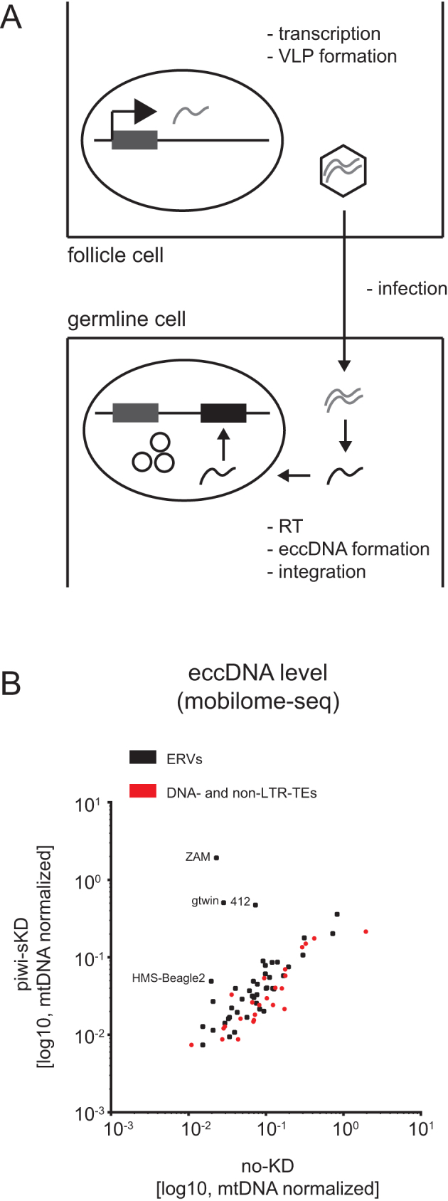
Figure 3.

Somatic piwi depletion may lead to ERV virus like particle (VLP) germline infection. (A) Schematic representation of ERV life cycle. ERV transcription, translation and VLP formation takes place in the somatic follicle cells. The VLP is then crossing by an unknown mechanism the cell-cell border between follicle cell and germ line cell. RNA is reverse transcribed into cDNA. Viral cDNA enters the germ cell nucleus and integrates. EccDNA is formed as a by-product of integration. (B) Scatter plot showing the number of TE mapping mobilome-seq reads, normalized to the number of mitochondrial DNA (mtDNA) mappers, in embryos after piwi-sKD or no-KD (log10 scale). ERVs are depicted as black dots, DNA- and non-LTR-TEs as red dots.
