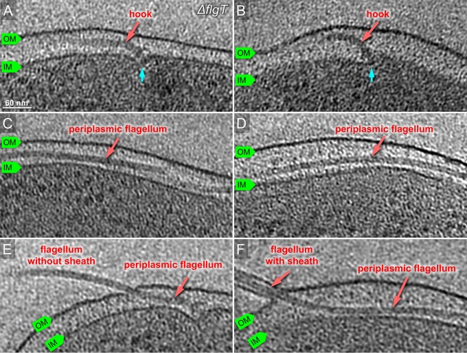
FIG 3.
Characterization of the ΔflgT flagellum in situ by cryo-ET. (A, B) Representative slices of tomograms from KK148 ΔflgT cells. The motor is visible beneath of outer membrane. The motor is indicated in cyan. (C, D) Representative slices of from KK148 ΔflgT cells. The flagellar filament is visible in the periplasmic space and labeled in red. (E) The flagellar filament is extended in the periplasmic space and penetrates the outer membrane without a sheath. (F) The flagellar filament penetrates from the periplasm and is sheathed.

