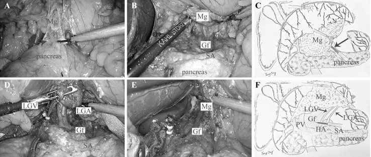
Fig. 3.
A The dissection began tracing the anterior pancreas space (arrow). B Dissect rearward following the surface of SA and expose the Gf. C The diagram of approach and separation space. D Dissection oriented by the Gf, and LGA and LGV were visible. E The LGA and LGV were both ligated at the root. F The diagram of rEME in the supra-pancreatic region. (Color figure online)

