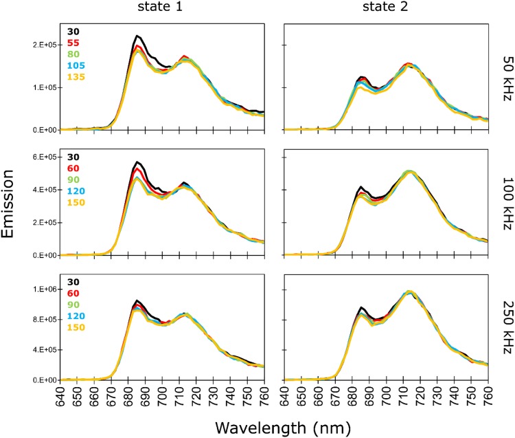
Fig. 2.
77 K steady-state fluorescence spectra reconstructed from globally analysed time-resolved fluorescence measured upon excitation at 400 nm in C. reinhardtii WT after incubation for 45 min under St1 conditions (left panel) or under St2 conditions (right panel). The spectra were normalized using a scaling factor obtained upon global analysis (more details in Materials and Methods). For each laser repetition rate, state 1 and state 2 spectra have the same vertical axis, and different colours of the spectra represent different cumulative exposure energies indicated in mJ (Fig. 1)

