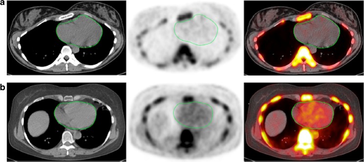
Fig. 7.
Transverse images (left CT, middle PET, right PET/CT) of the heart (green circles) in two clinically normal subjects (a 25 years old, b 61 years old). The global cardiac calcification scores were 12,492.44 in subject a and 18,424.70 in subject b. Normalizing the values to background NaF uptake increases the discrepancy between the subjects, resulting in 2.18 times the uptake in subject b than in subject a. Corresponding to the sites of NaF uptake in subject b, no structural calcification is seen on the corresponding CT scan and there is significant disparity between the PET and CT results. This is not an uncommon observation in this setting and clearly demonstrates the basis for assessment of cardiovascular calcification with these two different imaging modalities. While molecular imaging with NaF detects the earliest evidence for vascular calcification, evidence for calcification on CT largely reflects an end-stage disease process and therefore may be an irreversible pathologic state. Disparity between these two observations provides evidence for stage of calcification and has implications for the irreversibility of macrocalcification

