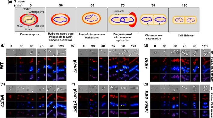
Figure 2.

Microscopic analysis of chromosome replication in outgrowing spores of different B. subtilis strains. (a) Schematic representation of spore germination/outgrowth stages employed for microscopic analysis. See text for details. (b–g) Dormant spores of wild‐type (b), ∆uvrA (c), ∆mfd (d), ∆disA (e), ∆disA uvrA (f), and ∆disA mfd (g) strains, were heat shocked and germinated as described in Materials and Methods. At different times (0, 30, 60, 75, 90, and 120 min) during germination/outgrowth, cells were collected, fixed and analyzed by bright‐field (BF) and fluorescence (DAPI and FM4‐64 staining) microscopy as described in Materials and Methods. Overlain images of DAPI and FM4‐64 at each time point are depicted as MERGE. Scale bar, 5 μm. Yellow arrowheads show cells that have replicated (at 75 min) and segregated their chromosome (at 90 min). For each strain, >200 cells were analyzed in at least six different fields
