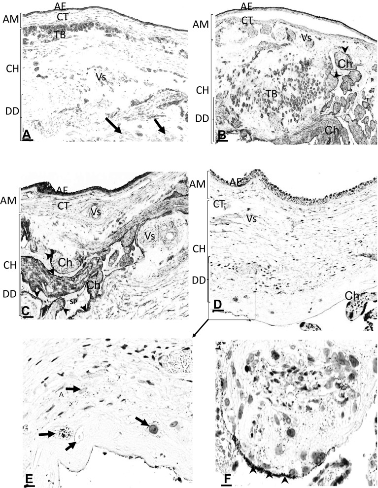
Fig. 2.
Location of AnxA2, S100A10 and the AnxA2/S100A10 complex in the foetal part and chorionic plate of the placenta. Sections from the same placental block were stained for AnxA2 (a, b), S100A10 (c) or the AnxA2/S100A10 complex (d–f). Representative images of the foetal side of the placenta are shown in low magnification. Amniotic (AM), chorionic (CH) and decidual (DD) parts are indicated, including the amniotic epithelium (AE), amniotic connective tissue (CT), chorionic trophoblasts (TB), chorionic villi (Ch), decidual cells (arrows in a), syncytiotrophoblast layers (arrowheads in b, c and f) and spiral vessels (SP in c). e, f show higher magnifications of proximity ligation stained sections indicating expression of the complex in the trophoblasts (arrows; e) and at the marginal surface of the syncytiotrophoblast (arrowheads; f). Scale bars: a, b 70 µm, c 60 µm, d 50 µm, e 20 µm, f 10 µm. Staining is as described in the legend of Fig. 1

