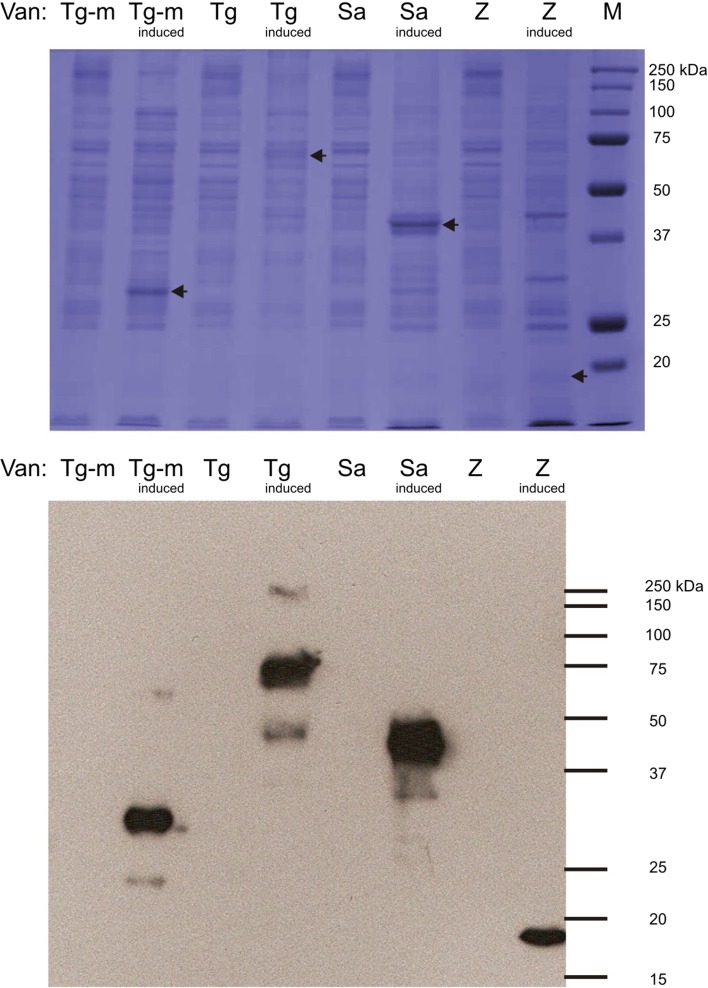
Fig. 1.
Expression in E. coli BL21 [DE3] mixed membranes of intact His6-tagged vancomycin resistance proteins: VanTG (serine racemace) of E. faecalis BM4518, VanTG-M (residues 1–342; a truncated version of native VanTG encoding only the putative serine membrane transporter portion), VanSA and VanZA of E. faecium B4147. Mixed membranes (10 μg) from cultures harbouring pTTQ-vanTG, pTTQ-vanTG-M, pTTQ-vanSA (Phillips-Jones et al. 2017a) or pMR2-vanZ (Rahman et al. 2007) expression plasmids and induced with 1 mM iso-propylthiogalactoside (IPTG) or uninduced cultures carrying the same plasmids were loaded on SDS-polyacrylamide gels (4% stacking/12% resolving). Following electrophoresis, gels were either stained with Coomassie Brilliant Blue for visual detection of protein bands (uppermost panel) or proteins were transferred electrophoretically onto PVDF membrane for Western blotting using an INDIA His probe followed by exposure to photographic film (lowermost panel)

