
Fig. 4.
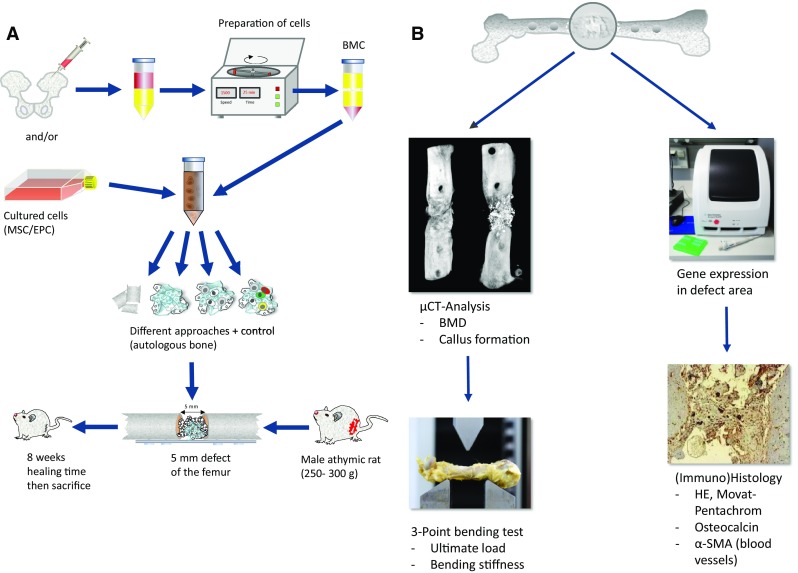
General scheme of the experimental setup to test various human cell types or scaffolds regarding their effect on bone healing is depicted in (a). The analyses made to evaluate the bone-healing response consist of µCT analysis to evaluate BMD and architecture of the new formed bone in the defect area, the same samples will be then used to determine the mechanical strength of the defect site using the three-point bending test. Additionally, RT-PCR to analyze the expression of genes involved in bone repair is performed using small samples from the defect site. Those bones were subsequently subjected to (immuno) histology to localize structures, cell types and protein expression in the bone defect (b)