
Fig. 2.
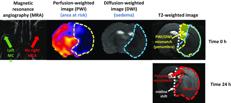
MRI images of a rat subject to middle cerebral artery (MCA) occlusion and reperfusion, illustrating recruitment of the ischaemic penumbra in the infarct. The top panels confirm the complete occlusion of the MCA and show the perfusion-weighted and diffusion-weighted images, which when combined reveal the ischaemic penumbra. In the lower panel, after 24 h part of the penumbra has been recruited into the area of infarct, and brain swelling has caused a quantifiable shift of the midline