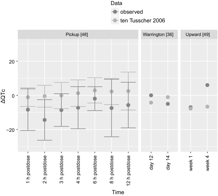
Fig. 5.
The results of PBPK-QSTS modeling in CSS in ten Tusscher and Panfilov [26] ventricular cardiomyocyte cell model (in blue) compared to clinically observed values (in red) of three clinical trials [36, 48, 49]. The results are presented as mean with standard deviation of ∆QTc (Color figure online)

