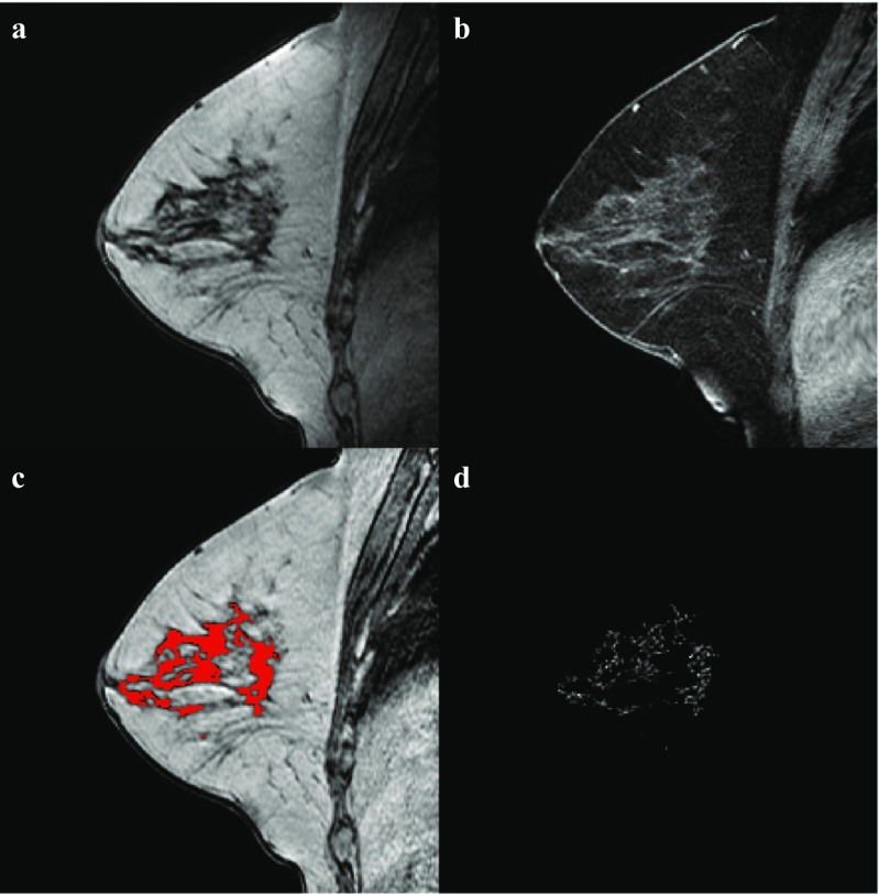
Fig. 1.
Example of the image processing in the contralateral breast of a 56-year-old patient with ER-positive/HER2-negative cancer patient. a non-fat-suppressed T1-weighted MRI, b fat-suppressed T1-weighted MRI after intravenous administration of contrast, c bias-field corrected image with the parenchymal tissue segmentation overlayed in red, d late enhancement in the parenchymal tissue segmentation

