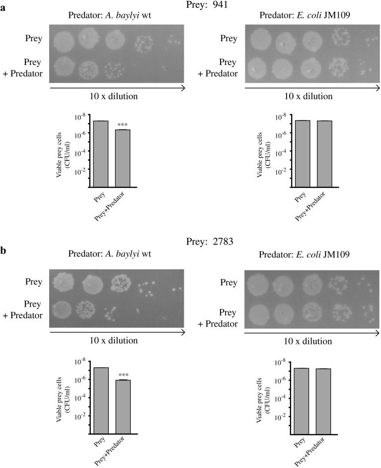
Fig. 2.
Growth competition assays. Δ941 (a) and Δ2783 (b) cells were incubated 4 h at 30 °C alone or mixed with a 10-fold excess of either A. baylyi ADP1 or E. coli JM109 cells before plating (see “Materials and Methods” for details). Images correspond to one representative experiment from three independent assays done with different cultures of prey and predator cells

