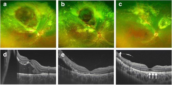
Fig. 2.
Preoperative and postoperative fundus photographs and optical coherence tomography images. Just before photodynamic therapy (PDT), subretinal exudation and haemorrhage were increased compared to the initial examination (a), and swept source optical coherence tomography showed the existence of macular detachment (d). At 1 month after PDT, the exudative retinal change had partially regressed (b and e). Although the subfoveal fluid had disappeared, the ellipsoid zone (Ez) was discontinuous and BCVA was 20/200 (e). At 10 months after PDT, both the subretinal haemorrhage and the exudative retinal detachment had disappeared completely (c and f). The Ez was partially recovered at 10 months after treatment (arrows, f), and BCVA had improved to 20/20

