Fig. 3.

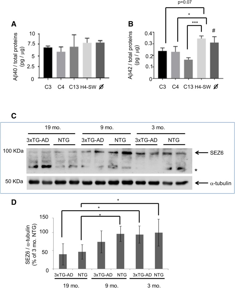
Evaluation of SEZ6 relevance for Alzheimer’s disease (AD) mechanisms in in vitro and in vivo models. a Quantification by enzyme-linked immunosorbent assay of soluble amyloid-β 1–40 (Aβ1–40) in conditioned media from H4-SW clonal lines (C3, C4, and C13) overexpressing SEZ6(R615H). The amyloid peptide concentration was normalized to the total protein content of the producing cells of each replicate. Measures are the mean ± SD of three independent wells. H4-SW Untransfected control; Ø H4-SW control transfected with pCDNA3.1 empty vector. b Same as in (a) except for the assessment of Aβ1–42 soluble form. * p < 0.05; *** p < 0.001, one-way analysis of variance (ANOVA) and post hoc test; # p < 0.05 vs. C4 and p < 0.01 vs. C13, one-way ANOVA and post hoc test. c Representative Western blotting for Sez6 protein detection in brain cortical extract from 3xTG-AD mice. Mice were killed at ages 3, 9, or 19 months, and Sez6 expression was assessed in transgenic and matched nontransgenic (NTG) animals. Each group was composed of three mice, and every animal was loaded in duplicate in the SDS-PAGE experiment. * Unspecific signal. d Densitometric quantification of all Western blot analysis data for Sez6 protein cortical expression (n = 3 mice/group) using ImageJ software. Each signal was normalized to the corresponding α-tubulin band to control for unequal protein loading. Results are expressed as a percentage of the youngest group (3 months) * p < 0.05, one-way ANOVA and post hoc test. mo. Months from birth
