Figure 1.

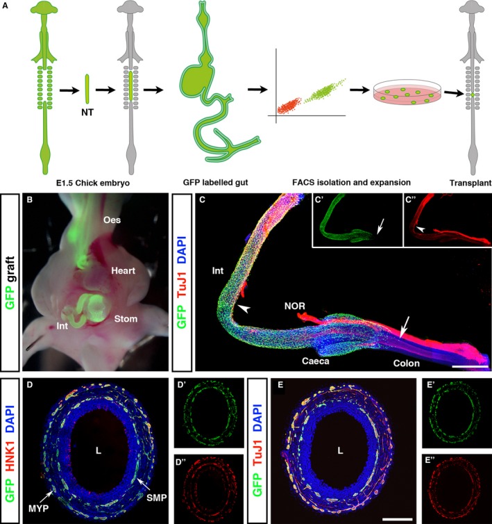
GFP chick intraspecies grafting efficiently labels the enteric nervous system of the gastrointestinal tract. (A) Schematic representing chick embryo tissue grafting methodology. Neural tubes from the vagal region (adjacent to somites 1–7) of GFP + E1.5 embryos were isolated and grafted into WT hosts in which the corresponding neural tube region was microsurgically ablated. Embryos developed for a further 12.5 days, at which point the intestines were harvested, stripped of mesentery, and dissociated into single cell suspension. GFP + cells were isolated by FACS and expanded in culture to form GFP + neurospheres for transplantation into E1.5 embryos. (B) Vagal neural tube grafting specifically labeled neural crest‐derived tissues including the ENS of the GI tract. (C) At E6.5, GFP + cells were observed along the gut, extending caudal to the caeca into the colon (C, arrow and C’). Following the migration wavefront, TuJ1+/GFP + cells were present (C, arrowhead and C’’). (D, E) At E8.5, the GI tract was completely colonised by migrating neural crest cells. Transverse sections of the colon revealed the formation of GFP + myenteric and submucosal plexuses. GFP + cells co‐expressed the neural crest cell marker HNK‐1 (D’’) and the neuronal marker TuJ1 (E’’). NOR, nerve of Remak. Scale bars: (C) 500 μm, (D, E) 250 μm.
