Figure 2.

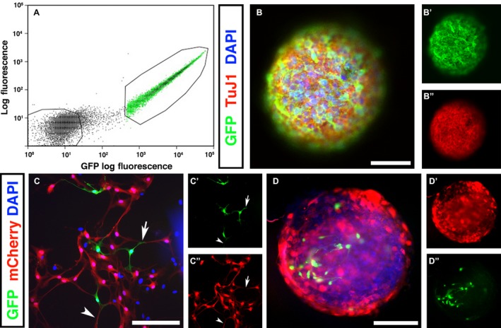
GFP + enteric neural crest‐derived cells form neurospheres and interconnections with CNS‐derived cells in vitro. After GFP tissue grafting, embryos were harvested at E14 and the gastrointestinal tract removed. (A) Gut tissues were dissociated into single cell suspension and sorted based on GFP expression and size. GFP –populations were collected and cultured as controls. (B) Following FACS isolation, GFP + graft‐derived cells formed free‐floating neurospheres after 1–2 weeks in culture. The majority of cells within neurospheres were immunopositive for the neuronal marker TuJ1 (B’). (C) GFP + neural crest‐derived cells were co‐cultured with spinal cord (SC)‐derived cells (labelled with an mCherry lentiviral construct). C’ and C’’ show higher magnification selections of (C). (D) After several days in culture, SC‐derived cells (red, D’) and ENS‐derived GFP + cells (D’’) aggregated to form mixed‐population neurospheres. Scale bars: (B, C) 50 μm, (D) 100 μm.
