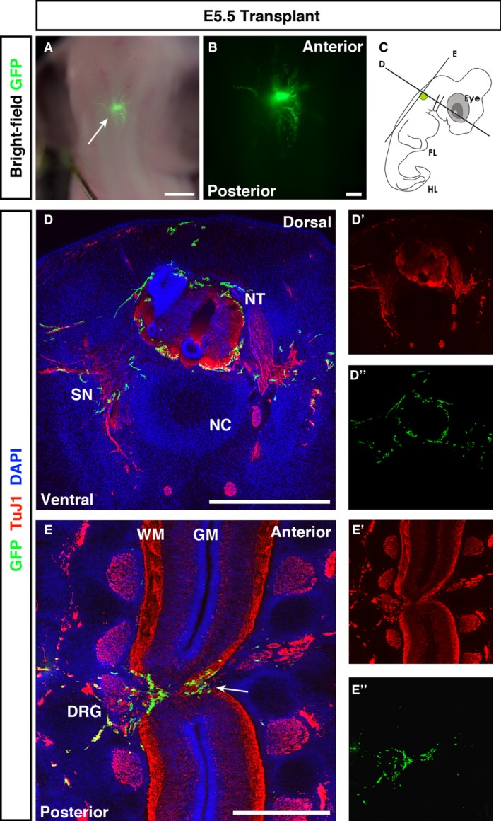
Figure 4.

E5.5 transplanted embryos show spread of GFP + cells through the white matter of the spinal cord. (A, B) Fluorescent stereoscopic examination revealed spread of GFP + ENSC from the transplantation site. (C) Schematic of the transplantation site and transverse and longitudinal sectioning planes used for analysis. (D) Co‐staining of transverse sections with GFP and TuJ1 revealed transplanted ENSC in neuron‐rich regions. (E) In longitudinal sections, transplanted ENSC formed bridging connections through the injury zone, between the anterior and posterior spinal cord tissue (E, arrow). In both transverse and longitudinal sections, GFP + ENSC spread into the PNS through dorsal root ganglia (DRG; E). Numerous GFP + projections extended from the transplanted neurosphere. DRG, dorsal root ganglia; FL, forelimb; GM, grey matter; HL, hindlimb; NC, notochord; NT, neural tube; SN, spinal nerve; WM, white matter. Scale bars: (A) 3 mm, (B) 1 mm, (D, E) 500 μm.
