Figure 7.

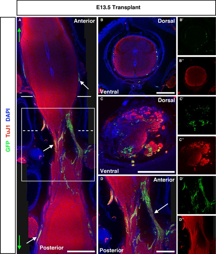
E13.5‐transplanted embryos show extensive bridging of GFP + ENSC across the injury site and substantial anterior/posterior spread. (A) Tiled images of longitudinally sectioned E13.5 embryos revealed the extent of ENSC spread along the anterior/posterior axis (maximum spread indicated by green arrows, white arrows highlight GFP + cells). Solid and dashed lines in (A) show the approximate plane of transverse sections shown in (B) and (C), respectively, and the solid box indicates the higher magnification of the injury site shown in (D). (B) Coronal section of the transplanted SC rostral to the transplantation site shows few GFP cells localised to the SC periphery, and some spread into the PNS. (C) Coronal sections within the injury zone reveal GFP + cells within both white and grey matter. (D) Higher magnification of the injury zone demonstrates the extensive formation of GFP + bridging strictures between the anterior and posterior SC across the injury zone. Scale bars: (A) 1000 μm; (B–D) 500 μm.
