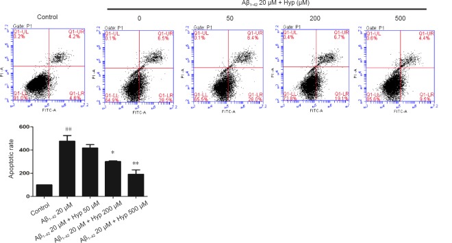
Figure 2.
Protection of Hyp against Aβ1–42-induced apoptosis in bEnd.3 cells as detected by flow cytometry (FITC-V/PI) analysis.
The apoptosis rate of fibrillar Aβ1–42-treated bEnd.3 cells obviously increased, and dramatically decreased after pretreatment with concentrations of Hyp from 50 to 500 µM. The results are presented as the mean ± SEM, and analyzed by one-way analysis of variance followed by Tukey's multiple comparison post hoc test. The experiment was conducted in triplicate. ##P < 0.01, vs. control group; *P < 0.05, **P < 0.01, vs. 20 µM Aβ1–42 group. Aβ1–42: Amyloid beta 1–42; Hyp: hyperoside.

