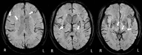
Figure 2.

Different sections of susceptibility-weighted images showing CMBs in the FSC region.
A 61-year-old man with a 6-year history of hypertension had CSVD-V-CIND at baseline and had developed dementia at the 1-year follow-up. CMBs (white arrows) in the FSC regions were defined as CMBs within the frontal lobe, frontal white matter, thalamus, and basal ganglion. The total number of FSC CMBs in this patient was 21. The three images represent different MRI sections of the brain. CMBs: Cerebral microbleeds; FSC: fronto-subcortical circuits; CSVD: cerebral small vessel disease; V-CIND: vascular cognitive impairment, but no dementia; R: right; L: left.
