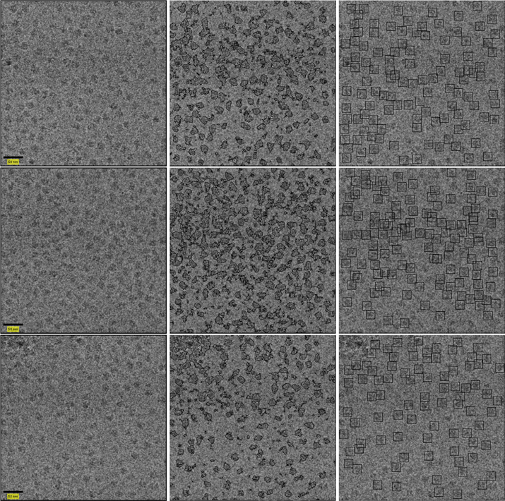
Fig. 12.
Picked particles of sample 70S ribosome micrographs without CTF correction. The micrographs are presented in the left column. Classification results are presented in the center. The picked particles are on the right. Defocus values are 1671.8, 1643.2 and 1595.6 nm for the top, middle and bottom micrographs, respectively.

