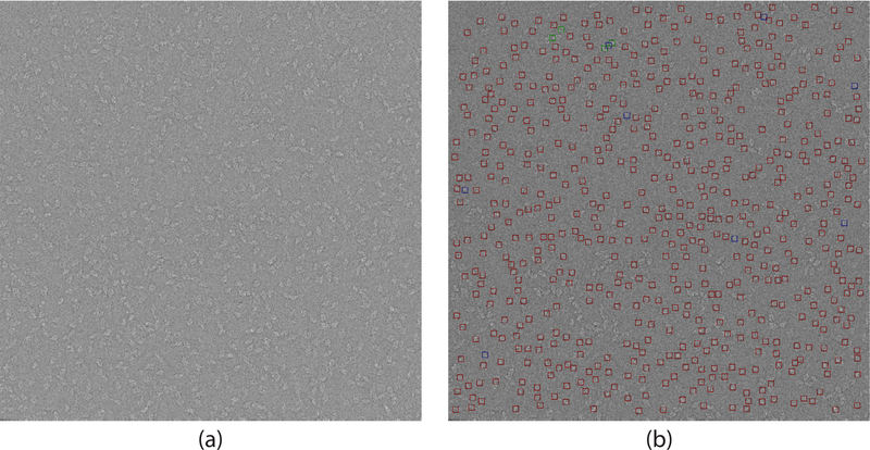
Fig. 6.
Illustration of picking with and without CTF correction. (a) A CTF-corrected β-galactosidase micrograph. CTF estimation was done using CTFFIND4 (Rohou and Grigorieff, 2015) followed by phase-flipping. (b) Results. Particles picked in both micrographs are surrounded by a red box. Selections unique to the phaseflipped micrograph are surrounded by a green box. Selections unique to the original micrograph are surrounded by a blue box. Defocus value for the micrograph is 4191.1 nm.

