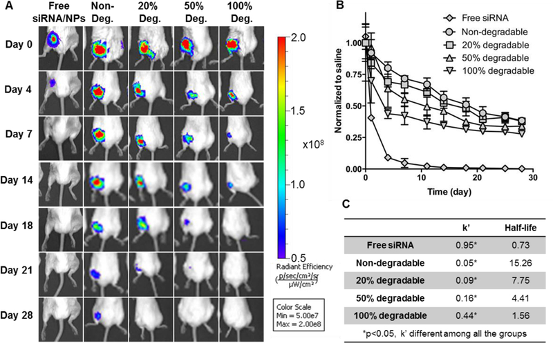
Fig. 5.

In vivo siRNA-NP localization qualitatively decreased as the hydrogel degradability increased as visualized by MS fluorescent imaging. (A) Representative IVIS images of Cy5- siRNA/NP loaded hydrogels placed around femur fractures over time. (B) Quantification of siRNA/NP localization from IVIS imaging. Data showed the total radiant efficiency of drawn region of interest (ROI) in IVIS images normalized to day 0. Mean ± STDEV, N=6. (C) Rate constant (k’) and half-lives are derived from a one phase decay model.
