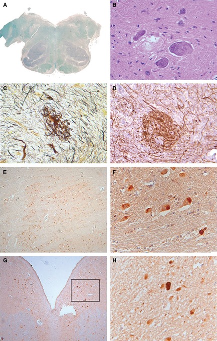
Figure 1.

(A–D) Show staining of the IOs. (A) Luxol fast blue stain shows symmetrically enlarged IOs. (B) H&E stain shows filamentous bundled cytoplasmic inclusions in IO neurons. Many neuronal cells show prominent nuclei containing granular nucleoplasm and large nucleoli. (C) Glomeruloid bodies are found in IOs (Bielschowsky silver stain). (D) IHC with anti‐200‐kDa neurofilament protein in hypertrophic processes. (E–H) Pontine 4R tau and 3R tau immunoreactivity. (E and F) 4R tau immunoreactivity. (G and H) 3R tau immunoreactivity.
