Figure 2.

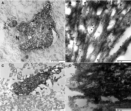
Immunolabeling of tau filaments using PHF1 antibody and ABC method plus silver enhancement (see an earlier report43 for the methodology). A neuron (A and B) and a neurite (C and D) granule are shown. N, nucleus.

Immunolabeling of tau filaments using PHF1 antibody and ABC method plus silver enhancement (see an earlier report43 for the methodology). A neuron (A and B) and a neurite (C and D) granule are shown. N, nucleus.