Figure 1.
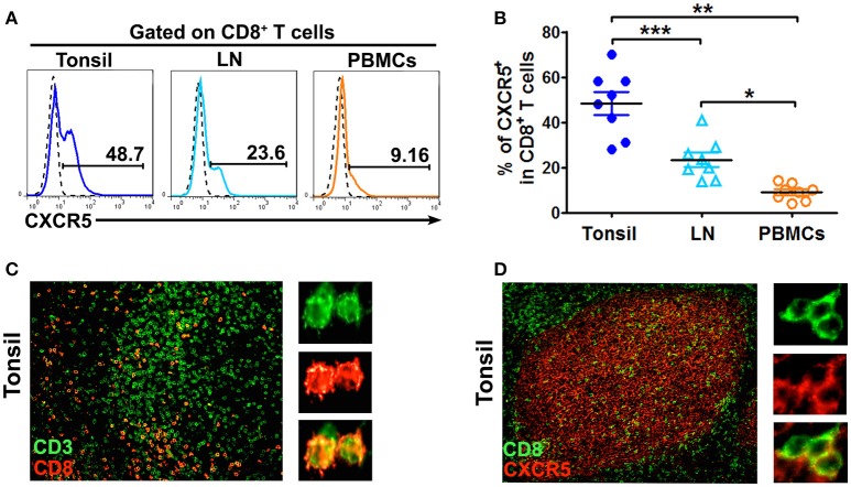
CD8+ T cells localized in B cell follicles in tonsils and lymph nodes express CXCR5. The expression of CXCR5 on CD8 T cells in tonsils, lymph nodes and PBMCs was shown in the representative histogram graphs (A) and summary data (B, n = 8). Immunofluorescence staining of CD3+ T cells (green) and CD8+ T cells (red) (C, n = 5), CD8+ T cells (green) and CXCR5 (red) (D, n = 5) in paraffin-embedded tonsil tissues. Data are expressed as the mean ± SD, and compared with Mann-Whitney test. *P < 0.05, **P < 0.01, and ***P < 0.001.

