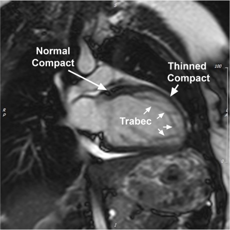
Fig. 2.

Magnetic resonance image (2-chamber view) of mild dilated cardiomyopathy with noncompaction left ventricle (LV) shows the LV during end-diastole. Note the typical structural changes of the thinned compact layer at the level of noncompaction architecture (“thinned compact”) in comparison with the normal LV segment (“normal compact”). This individual has morphologic signs of noncompaction for 50% of the LV circumference (indicated by a line in the accompanying motion image).
Trabec = trabeculations
Supplemental motion image is available for Figure 2 (3.4MB, mp4) .
