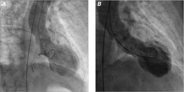
Fig. 2.

Patient 1. On A) second and B) third presentation, left ventriculograms (right anterior oblique views) during systole show regional wall-motion abnormalities consistent with a midventricular variant of takotsubo cardiomyopathy.
Supplemental motion images are available for Figure 2A (3.2MB, mp4) and 2B (3.2MB, mp4) .
