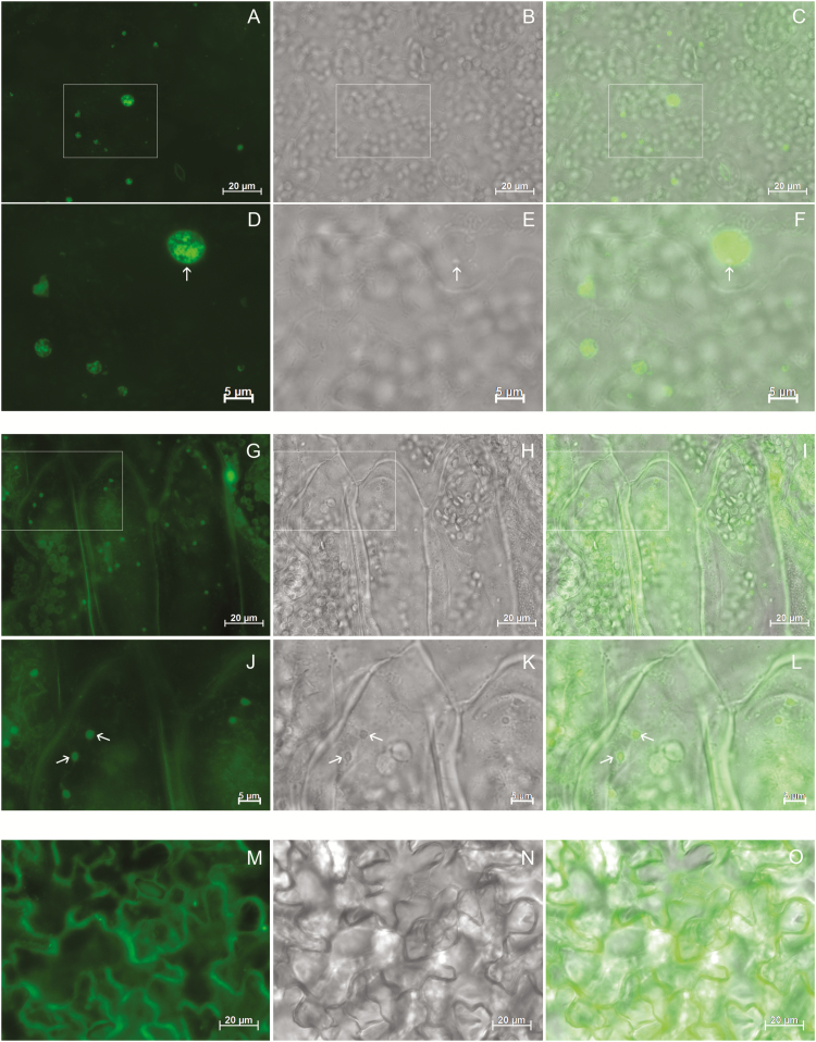
Fig. 8.
27γzf protein bodies (PBs) and 16γzf endoplasmic reticulum (ER) enlargements can be detected by fluorescence staining of the ER. Leaf tissue from 16γzf (A–F), 27γzf (G–L) or wild-type (M–O) Arabidopsis plants was stained with DiOC6 dye and examined using epifluorescence microscopy. (A, D, G, J, M): DiOC6 fluorescence (green); (B, E, H, K, N): bright-field; (C, F, I, L, O): merged images. Camera exposure time (ms): 61 (A), 502 (G), 8352 (M). Boxes in (A–C) and (G–I) indicate the regions that are shown at higher magnification in (D–F) and (J–L), respectively. Arrows indicate enlarged ER (D–F) or PBs (J–L).

