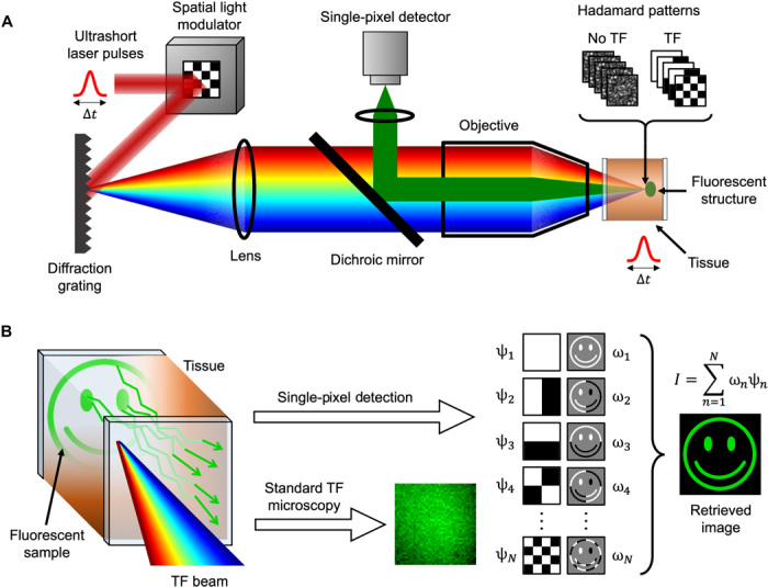
Fig. 1. Working principle of TRAFIX.

(A) A femtosecond laser beam is expanded onto a spatial light modulator (SLM) that generates Hadamard patterns. Subsequently, the beam is diffracted from a grating, and the Hadamard patterns are projected onto a fluorescent sample after propagating through a scattering medium. Fluorescent light emitted by the sample is collected by the same objective after passing through the scattering medium a second time (epifluorescence geometry), and the total intensity is measured by a single-pixel detector. (B) A TF beam propagates through a turbid medium with minimal distortion, retaining the integrity of illumination patterns in the sample plane. Emitted fluorescent photons scatter as they propagate back through the tissue. In contrast to standard TF microscopy, TRAFIX tolerates scrambling of back-propagating light since only an intensity measurement is performed. In a single-pixel measurement, the fluorescent target is sequentially illuminated with Hadamard patterns (ψn), and the total intensity detected is stored as a coefficient (ωn). Gray background in the second column denotes regions of zero intensity. By adding up the Hadamard patterns weighted by their respective coefficients, an image of the fluorescent sample is reconstructed.
