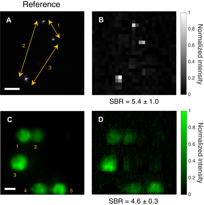
Fig. 3. Images of fluorescent microscopic samples through unfixed human colon tissue.

Fluorescent beads of 400 nm in diameter and fixed HEK293T/17-GFP cells were imaged through 250 and 200 μm of human colon tissue, respectively. (A and C) Images taken from the reference imaging system under uniform TF illumination across the FOV. Camera binning in (A) was set to 4 × 4, and exposure time was 5 s. No camera binning was used in (C), and exposure time was 15 s. (B and D) Images obtained with TRAFIX using a Hadamard basis containing 1024 and 4096 illumination patterns, respectively. All patterns were used for image reconstruction (CR = 1). Camera binning for each Hadamard pattern was set to 64 × 64, and exposure time values were (B) 1 s and (D) 0.75 s. The spacing between beads and the diameter of cells were measured to assess image quality (tables S3 and S2, respectively). The SBR is shown for all reconstructed images. Scale bars, 10 μm.
