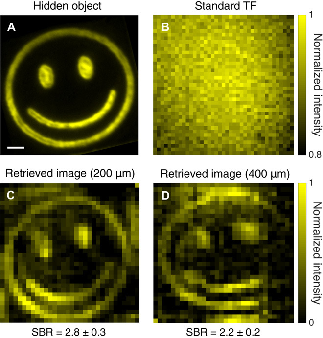
Fig. 4. Comparison of a hidden object and the retrieved images through fixed rat brain tissue.

(A) Reference image of a fluorescent micropattern without any scattering sample. (B) Image obtained by conventional TF microscopy (i.e., under uniform wide-field TF illumination with wide-field detection in epifluorescence configuration) through 400 μm of fixed rat brain tissue. (C and D) Reconstructed images obtained with TRAFIX through 200 and 400 μm of rat brain tissue, respectively. The two retrieved images were reconstructed using a full Hadamard basis containing 1024 patterns. Camera binning was set to 64 × 64, and exposure time values were (C) 0.2 s and (D) 1 s. Small intensity variations in the reconstructed images arise from inhomogeneities in the fluorescent micropattern originated in the imprinting process. Larger intensity variations are due to inhomogeneities in light transmission through the highly anisotropic scattering medium. This also applies to figs. S7, S9, S10, and S12. The SBR is shown for all reconstructed images. Scale bar, 10 μm.
