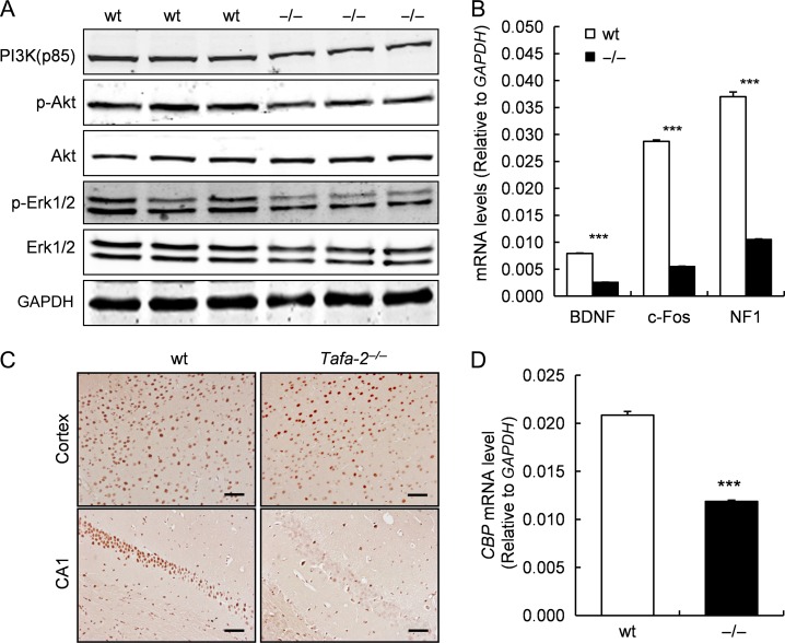
Figure 8.
Downregulaton of PI3K/Akt and MAPK/Erk signaling pathways and reduced expressions of CREB-dependent genes and CBP in the brain of Tafa-2−/− mice (A) Western blot analysis of PI3K (p85), p-Akt, Akt, p-Erk1/2, Erk1/2 in the brain lysates of wt and Tafa-2−/− mice. GAPDH was used as a protein loading control. (B) Real-time PCR analysis of BDNF, c-Fos, and NF1 mRNA levels in the brain of wt and Tafa-2−/− mice. GAPDH was used as an internal control. (C) Representative images of immunohistochemistry of CBP on sections of cortex and hippocampus CA1 from wt and Tafa-2−/− mice. Scale bar = 50 μm. (D) Real-time PCR analysis of CBP mRNA level in the brain of wt and Tafa-2−/− mice. GAPDH was used as an internal control. Data are shown as the mean ± SE. Student’s t test: ***P < 0.001, n = 3 for each genotype.

