Figure 2.
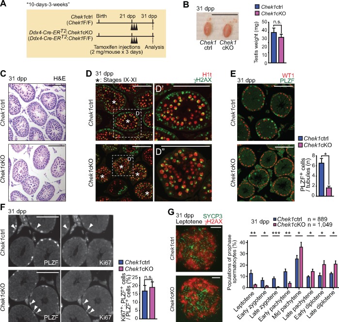
CHEK1 is required for the maintenance of spermatogonia. (A) Schematic of the experimental design of the 10-day-3-weeks method. (B) Picture of Chek1ctrl and Chek1cKO testes at 31 dpp. Scale bar: 1 cm. Testis weights are shown as mean ± SEM for three independent pairs of Chek1ctrl and Chek1cKO mice. n.s.: not significant. Significance was determined utilizing an unpaired t-test. (C) Sections stained with H & E at 31 dpp. Scale bars: 100 μm. (D) Immunostaining of H1t and γH2AX on testicular sections at 31 dpp. Asterisks indicate seminiferous tubules at stages IX–XI. Dashed squares are magnified in the right-hand panels. Scale bars: 100 μm. (E) Immunostaining of WT1 and PLZF on testicular sections at 31 dpp. Scale bars: 100 μm. Numbers of PLZF positive cells per seminiferous tubule are shown as mean ± SEM for three independent pairs of Chek1ctrl and Chek1cKO mice. *P < 0.05 (unpaired t-test). (F) Immunostaining of PLZF and Ki-67 on testicular sections at 31 dpp. Arrowheads indicate cells that are double positive for PLZF and Ki-67. Scale bars: 100 μm. Percentages of PLZF-positive cells that are also positive for Ki-67 are shown as the mean ± SEM for three independent pairs Chek1ctrl and Chek1cKO mice. n.s.: not significant. Unpaired t-test. (G) Immunostaining of SYCP3 and γH2AX on meiotic chromosome spreads at 31 dpp. Scale bars: 10 μm. Stage populations during meiotic prophase are shown as the mean ± SEM for five independent pairs of Chek1ctrl and Chek1cKO mice. Total numbers of analyzed nuclei are indicated in the panels. Scale bars: 10 μm. *P < 0.05, **P< 0.01, ***P < 0.001 (unpaired t-test).

