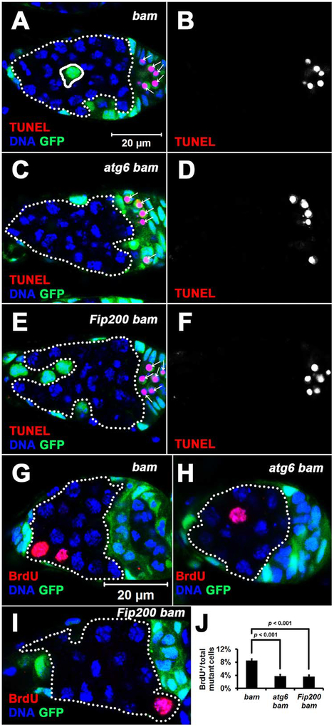
Figure 5. Cell Death Is Not Altered, but the Cell Cycle Is Attenuated in Autophagy-defective bam Mutant Cells.
(A-F) Cell death is not altered in autophagy-defective bam mutant cells. Homozygous bamΔ86, atg6Δ1 bamΔ86, and Fip2003F5 bamΔ86 mutant cells (GFP− cells outlined by white dotted lines) were analyzed at 14 days after the last heat shock. bamΔ86, atg6Δ1 and Fip2003F5 are all null alleles. White arrows denote dying neighboring follicle cells. All images are the same magnification.
(G-J) The cell cycle is attenuated in autophagy-defective bam mutant cells. (G-I) Homozygous bamΔ86, atg6Δ1 bamΔ86 and Fip2003F5 bamΔ86 mutant cells (GFP− cells outlined by white dotted lines) were analyzed at 7 days after the last heat shock. Images are the same magnification. (J) Quantification of BrdU incorporation in homozygous bamΔ86, atg6Δ1 bamΔ86 and Fip2003F5 bamΔ86 mutant cells. The mutant germ cells in single confocal microscope focal plane of 30 germaria were quantified for each replicate, and three independent replicates were performed for each genotype. Data represent mean ± standard deviation, and statistical significance was determined by a two-tailed Student’s t test for two samples assuming unequal variances.
See also Figure S4, S5, S6, and Tables S1 and S2.

