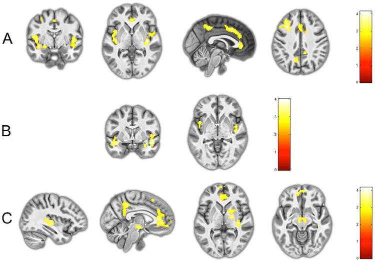
Figure 1.

a) Reduced activity in the amygdala (far left), insula (middle left), ACC and PCC (middle right), and dorsolateral prefrontal cortex (far right) in the Primary group compared to the Comparison group during fear processing; b) Trendwise reduced activity in the amygdala (left) and insula (right) in the Primary compared to Secondary group during fear processing; c) Reduced activity in the superior temporal sulcus/inferior parietal lobe (far left), ACC and PCC (middle left), thalamus and globus pallidus (middle right), and susbtantia nigra (far right) in Secondary group compared to the Comparison group during fear processing.
